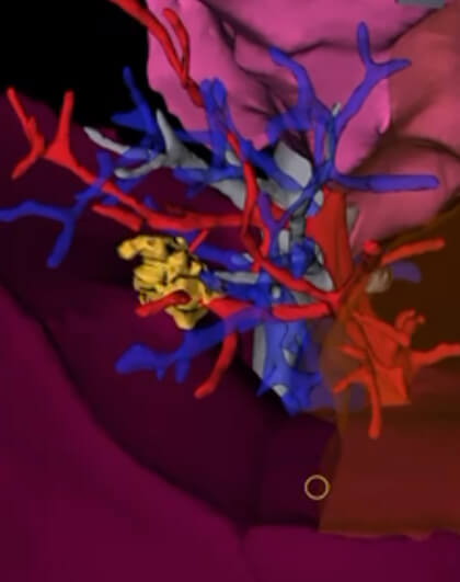
Since November, Emory surgeons have been piloting new software that creates a three-dimensional digital image of a patient's lungs. Having access to a 3D model makes removing hard-to-reach cancerous cells more efficient and precise. Think of it as the first time a cartographer added mountains and canyons to the layout of a map — only in this case, that map leads straight to saving a patient's life.
At Emory Saint Joseph's Hospital, Manu Sancheti, MD, and his team are the first thoracic surgeons in the Southeast, and only the third in the country, to explore how this new Intuitive 3D Modeling tool can aid robotic-assisted thoracic surgery.
Non-small cell lung cancer, in particular — the kind that most benefits from robotic surgery and 3D imaging — can often be challenging to pinpoint and remove since the tissue samples can exist deep within the lung. With a combination of robotic-assisted surgery and these enhanced imaging techniques, it is quicker and easier to figure out how to approach suspicious-looking nodules.